„”

Wir befassen uns mit biologischen Fragen und nutzen das gesamte Repertoire an lichtmikroskopischen Techniken (konfokale Laser-Scanning-Mikroskopie, Zwei-Photonen-Anregung, Uncaging-Prozesse, FCS, FRAP, FRET, FLIM, zelluläre Aufnahme von Peptiden, Kolokalisationsstudien, intrazelluläre Ionenkonzentrationen, Proteinübersetzungen usw.) in Verbindung mit elektrophysiologischen Techniken.
Auf dieser Seite
Die Technologieplatform Zelluläre Bildgebung unterstützt Forschende an verschiedenen Stadien ihrer Mikroskopieprojekte von Projektdesign, über Probenvorbereitung, Geräteeinweisungen bis zu Bild-und Datenanlyse.
Lichtmikroskopie
Wir stellen Wissenschafltern des FMP und externen Kollaborationspartnern modernste Fluoreszenzmikroskope für Untersuchung an lebenden Zellen und Geweben zur Verfügung. Mittels Totaler Interner Reflektions - (TIRF) sowie Superauflösungsmikroskopie kann die Nanostruktur von Protein und Membrankomplexen dargestellt werden. Weitere Methoden für Ionenkonzentrationsmessungen, lichtgesteuerte Freisetzung von Pharmaka sowie zur Untersuchung von molekularen Interaktionen mittels FRET, FRAP, FLIM oder FCS werden angeboten.
Elektronenmikroskopie
Svea und Dima bieten Wissenschafltern des FMP und externen Kollaborationspartnern Präperationstechniken und modernste Elektronenmikroskope zur Darstellung zellulärer Ultrastrukturen und Lokalisierung von Proteinen an. Sie helfen Nutzern bei der Probenherstellung, Immunogoldmarkierung sowie Bildgebung und Analyse mittels 3D (Tomografie & FIB-SEM) und korrelativer Licht-und Elektronenmikroskopie.
Bildanalyse
Gayathri Nadar berät und trainiert Nutzer um mittels Bildanalyseprogrammen (ImageJ, Huygens, Imaris) quantitative Messungen aus Bilddaten zu generieren. Weiterhin werden neue, benutzerdefinierte Skripte und Programme entwickelt um Bilddaten darzustellen und zu analysieren. Alle zwei Wochen wird "Image Analysis Table Talk" angeboten, bei dem alle Nutzern vorbeikommen können, um über Bildanalyseprojekte zu sprechen und Hilfe zu erhalten.
STEDYCON
STEDYCON
Workstation
Workstation
Software
Software
Software
Software
Packages available
Workstation
Workstation
Software
Software
Software
Software
Packages available
Mit Hilfe von zellbiologischen und mikroskopischen Methoden untersuchen wir: (i) die Auswirkung der molekularen Zusammensetzung der Tight Junctions auf deren Barrierefunktion und Ionenselektivität, (ii) die Funktion von Mitochondrien mittels MitoMap, einem integrativen Modell basierend auf Cross-Linking-MS, Superauflösungs- und Elektronenmikroskopischer Bildgebung (in Zusammenarbeit mit Fan Liu) und (iii) Wirkmechanismen neuer Medikamente und toxischer Verbindungen mittels hochauflösender phänotypischer Hochdurchsatzmikroskopie (in Zusammenarbeit mit Han Sun und Jens Peter von Kries).

![[Translate to Deutsch:] FAF2](/fileadmin/_processed_/4/2/csm_FAF2_Truncation11002_ed2b940128.png)
![[Translate to Deutsch:] EM1](/fileadmin/Data/Molecular_Physiology_Cell_Biology/Martin_Lehmann/Images/EM1.jpg)
![[Translate to Deutsch:] Embryo](/fileadmin/Data/Molecular_Physiology_Cell_Biology/Martin_Lehmann/Images/ScientificImageoftheMonth_RozemarijnvanderVeen.png)
![[Translate to Deutsch:] Mitochondria](/fileadmin/Data/Molecular_Physiology_Cell_Biology/Martin_Lehmann/Images/IMG0019_A2_1uM_BG-SiR_d12_live_HP_coverslip_ROI01.jpg)
![[Translate to Deutsch:] EM2](/fileadmin/Data/Molecular_Physiology_Cell_Biology/Martin_Lehmann/Images/EM2.jpg)
![[Translate to Deutsch:] Meshwork](/fileadmin/Data/Molecular_Physiology_Cell_Biology/Martin_Lehmann/Images/IMG0067_Sample_name.png)

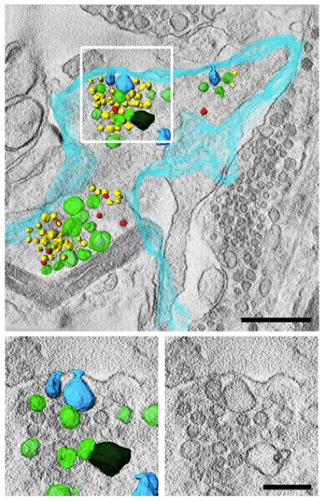
3D-Elektronenmikroskopaufnahmen von elektrisch stimulierten Synapsen, die mit VPS34IN1 behandelt wurden, zeigen multiple Einstülpungen der Plasmamembran (türkis), synaptische Vesikel (gelb) und endosomenähnliche Vakuolen (grün), Maßstabsbalken 500 nm (oben) und 200 nm (unten).
Liu, Guan-Ting et al. EMBO, May 2022
TMEM206 ist auf der MP-Membran vorhanden. MPs aus lebenden WT-BMDMs, die mit TMEM206-GFP (grün) transfiziert wurden, wurden mit 70 kDa TMR-Dextran (rot) nach M-CSF-Stimulation mittels Spinning-Disc-Mikroskopie bei 37 °C sichtbar gemacht. Maßstabsbalken, 5 µm. Bildaufnahme mit zwei Bildern pro Minute. Filmgeschwindigkeit, zwei Bilder pro Sekunde.
Zeziulia, M., Blin, S., Schmitt, F.W. et al. Nat Cell Biol, Mai 2022
FAF2 (magenta) weist, mithilfe von konfokaler Mikroskopie, eine duale Lokalisation sowohl in Mitochondrien als auch dem ER auf, anhand der Co-Lokalisierung mit TOM20 (cyan) als Marker für die äußere Mitochondrienmembran sowie Calreticulin (gelb) als Marker für das endoplasmatische Retikulum.
Elektronenmikroskopische Aufnahme einer Betazelle in der Bauchspeicheldrüse
Färbung der Tight Junction Proteine Occludin (grün) und Claudin-3 (rot), kombiniert mit einer Kernfärbung (DAPI, blau) in einem Mausembryo im letzten Stadium vor der Geburt (E18.5)
Mitochondriale Cristae, visualisiert mittels STED-Mikroskopie in HeLa-Zellen
Elektronenmikroskopische Aufnahme einer Synapse im Bulbus olfactorius
Claudin-1 Netzwerkarchitektur in lebenden COS7 Zellen, visualisiert mittels STED-Mikroskopie
Der zellpermeable Lamin-Nanokörper in der STED-Mikroskopie. a) Schematische Darstellung des mit Atto594 markierten Anti-Lamin-Nanokörpers, der an die Kernlamina bindet. b) STED-Mikroskopie von HeLa-Kyoto-Zellen, die mit 2 μm des zellpermeablen Nanokörpers mit 500 nm SiR-Hoechst behandelt wurden. Maßstabsbalken 5 μm. c) Histogramm der normalisierten Fluoreszenzintensität über einer Linien-ROI (siehe weißer Kasten in (b)). Schneider, Anselm F L et ...
Das Leibniz-Forschungsinstitut für Molekulare Pharmakologie (FMP) gehört zum Forschungsverbund Berlin e.V. (FVB), einem Zusammenschluss von sieben natur-, lebens- und umweltwissenschaftlichen Instituten in Berlin. Die Einrichtungen sind Mitglieder der Leibniz-Gemeinschaft.
Leibniz-Forschungsinstitut für Molekulare Pharmakologie im Forschungsverbund Berlin e.V. (FMP)
Campus Berlin-Buch
Robert-Roessle-Str. 10
13125 Berlin, Deutschland


![[Translate to Deutsch:] background header](/fileadmin/_processed_/1/b/csm_Bacteriophage_final_v02_bb61a5ea72.jpg)

![[Translate to Deutsch:] link](/fileadmin/_processed_/0/6/csm_1_c223dcd589.jpg)

![[Translate to Deutsch:] background header](/fileadmin/_processed_/6/2/csm_forschergruppen_5_34300470f7.jpg)
![[Translate to Deutsch:] bachground picture](/fileadmin/_processed_/3/d/csm_Forschungsgruppen_ddee470de1.jpg)
![[Translate to Deutsch:] leibniz phd](/fileadmin/_processed_/3/f/csm_fmp-career-phd_aaf1698784.jpg)
![[Translate to Deutsch:] link](/fileadmin/_processed_/4/6/csm_Glaesernes_Labor-0032_3642a3cb69.jpg)
![[Translate to Deutsch:] Group photo](/fileadmin/_processed_/2/5/csm_IMG20220304141122_a89d728364.jpg)


![[Translate to Deutsch:] book](/fileadmin/_processed_/9/c/csm_book_fc96d87fa8.png)