„”

We address questions of biological and pharmacological importance using light microscopy techniques (confocal laser scanning microscopy, two-photon excitation, uncaging processes, FCS, FRAP, FRET, FLIM, cellular uptake of peptides, colocalization studies, intracellular ion concentrations, protein translations studies, etc.) in conjunction with the electron microscopic and electrophysiological techniques.
The Cellular Imaging Facility assists the users at every stage of their imaging projects, from project design, microscopy training, sample preparations, assistance in imaging to image data analysis and visualization.
Light Microscopy
The Light Microscopy Unit supports the researchers with high-end fluorescence imaging technology to study living cells, microorganisms, and tissue organization. We aid the study of proteins and membranes at the nanoscale using our super-resolution and TIRF microscopes. We also assist in single-cell and molecular imaging techniques such as FRET, FRAP, FLIM, TIRFM, FCS, ion measurements, and caged compounds.
Electron Microscopy
The Electron Microscopy Unit provides support in the visualization of cellular ultrastructure and in localizing individual proteins at the subcellular level. The EM unit also supports the users with sample preparation techniques, immunogold labeling, correlative light and electron microscopy (CLEM), 3D electron microscopy using tomograpical reconstruction or Focused Ion Beam Scanning Electron microsopy (FIB-SEM), and image data analysis.
Image Data Analysis
The Image Analyst in our facility provide consultation and training in image analysis methods and techniques for extracting meaningful quantitative measurements from images. We also support developing custom automated workflows for image data analysis and visualization. Support with image analysis software such as FIJI/ImageJ, Huygens, Imaris is also available. Every two weeks "Image Analysis Table Talk" is offered which is free for all users to come by and talk about image analysis projects and get help.

STEDYCON

STEDYCON

Workstation
Workstation
Software
Software
Software
Software
Packages available
Workstation
Workstation
Software
Software
Software
Software
Packages available
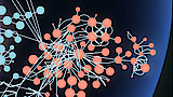
The central aim of our group is to establish suitable single cell techniques and the application of the most diverse microscopic methods to study the following processes: (i) Molecular composition, nanoscale organization and physiological function of tight junction as ionselective paracellular barrier (with Volker Haucke) (ii) Establish an integrated model of mitochondria based on cross-linking MS, Super-resolution and electron microscopy (with Fan Liu) (iii) Identification of toxic compounds and new pharmacological mechanisms via high-resolution confocal imaging (with Han Sun & Jens Peter von Kries).
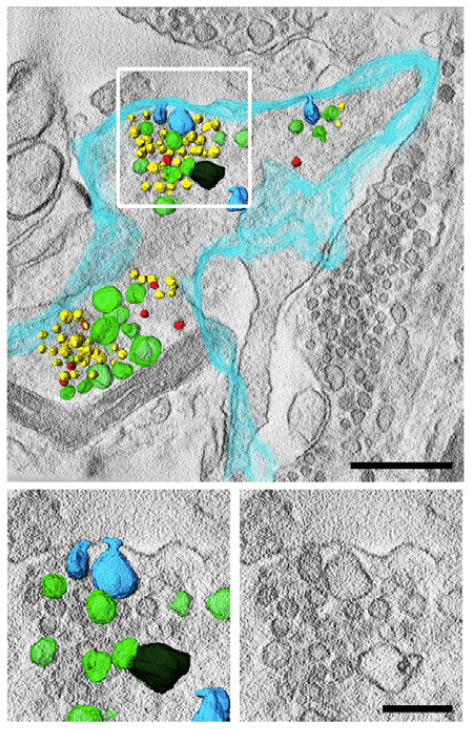


3D Electronmicrograph of electrically stimulated Synapses treated with VPS34IN1 show multiple invaginations from the plasma membrane (turquoise), synaptic vesicles (yellow) and endosome like vacuoles (green), scale bars 500 nm (top) and 200 nm (bottom).
Liu, Guan-Ting et al. EMBO, May 2022
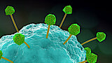
TMEM206 is present on MP membrane. MPs from living WT BMDMs transfected with TMEM206-GFP (green) were visualized with 70 kDa TMR–dextran (red) after M-CSF stimulation using spinning disc microscopy at 37 °C. Scale bar, 5 µm. Imaging of two frames per minute. Movie speed, two frames per second.
Zeziulia, M., Blin, S., Schmitt, F.W. et al. Nat Cell Biol, May 2022
FAF2 (magenta) shows dual-localization in confocal microscopy by co-localization with Tom20 (cyan) as a marker for the outer mitochondrial membrane as well as Calreticulin (yellow) as a marker for the endoplasmic reticulum.
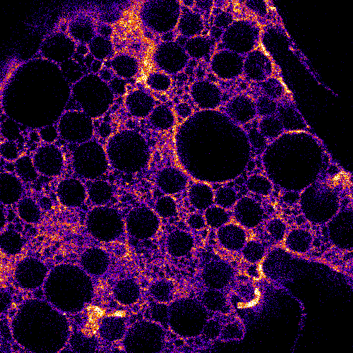
Electron microscopy image of a beta cell in the pancreas
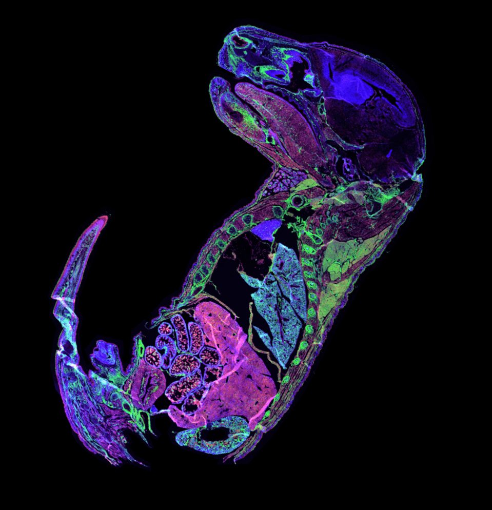
Staining of the Tight Junction proteins Occludin (green) and Claudin-3 (red), combined with a nuclear (DAPI, blue) staining in a mouse embryo in the last stage before birth (E18.5).
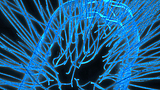
Mitochondrial cristae visualized using STED microscopy in HeLa cells
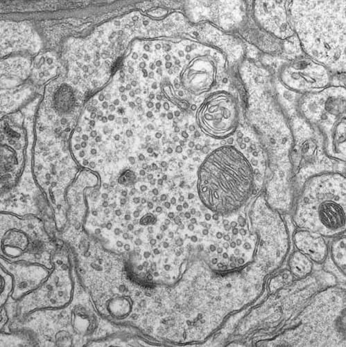
Electron microscopy image of a synapse in the olfactory bulb
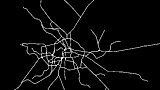
Claudin-1 meshwork architecture in living COS7 cells visualized with STED microscopy
The cell‐permeable lamin nanobody in STED microscopy. a) Schematic of the Atto594‐labeled anti‐lamin nanobody binding to the nuclear lamina. b) STED microscopy of HeLa Kyoto cells treated with 2 μm of the cell‐permeable nanobody with 500 nm SiR‐Hoechst. Scale bar 5 μm. c) Histogram of the normalized fluorescence intensity over a line ROI (see white box in (b)).
Schneider, Anselm F L et al. Angewandte Chemie (International ed. in English), Sep 2 ...

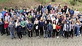
People
The Leibniz-Forschungsinstitut für Molekulare Pharmakologie (FMP) is part of the Forschungsverbund Berlin e.V. (FVB), which legally represents seven non-university research institutes - members of the Leibniz Association - in Berlin.
Leibniz-Forschungsinstitut für Molekulare Pharmakologie im Forschungsverbund Berlin e.V. (FMP)
Campus Berlin-Buch
Robert-Roessle-Str. 10,
13125 Berlin, Germany