„”
Methodological advances in DNA sequencing for patient diagnosis have enabled the identification of hundreds of variations in synaptic genes, associated with a broad spectrum of brain conditions. The overwhelming number of disease-related variations presents multiple challenges: Are all variations pathogenic? How do they affect synapse function? Is each effect unique? Or can converging processes be identified?
Our lab is studying disorders of the presynaptic terminal. We aim to identify mechanisms by which pathogenic variations affect synaptic function, and to develop strategies for fast diagnosis of variant pathogenicity. We believe that the teamwork of clinicians, geneticist and wet-lab researchers is essential to enable translation of knowledge from bedside to bench and back.
Our primary focus lies on a brain condition associated with genetic variation in a central synaptic protein called UNC13A (also known as Munc13-1). UNC13A is a major component of the presynaptic terminal, mediating a molecular preparation step of synaptic vesicles called ‘priming’. Priming enables synaptic vesicles to fuse with the plasma membrane in response to a signal in the transmitting neuron, release their neurotransmitter content, and thereby convey the signal to the receiving neurons. Dysfunction of UNC13A is bound to cause a signaling imbalance along the neuronal network. Genetic variations in UNC13A that are associated with disease were only recently identified, and little is known about the clinical and genetic spectrum of this condition, as well as on the cell biological and molecular mechanisms that underlie it. We believe that identifying the mechanism by which variations affect synaptic function will contribute to the development of therapeutic approaches to alleviate accompanying symptoms.
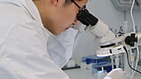
To assess how the properties of synaptic transmission are modulated by disease-related protein variations, we conduct whole-cell voltage clamp electrophysiological recordings in single, autaptic neurons in culture (Lipstein et al., JCI 2017). This experimental model enables an exquisite and in-depth readout of cell-autonomous processes. We complement our functional studies with confocal- and super-resolution microscopy analyses and with mass-spectrometric approaches, aiming to achieve a comprehensive molecular-functional link of diseased synapses.
More about the condition and its underlying molecular mechanisms can be found under https://www.jci.org/articles/view/90259
We encourage clinicians, geneticist, and patient families to contact us for further details.
The Leibniz-Forschungsinstitut für Molekulare Pharmakologie (FMP) is part of the Forschungsverbund Berlin e.V. (FVB), which legally represents seven non-university research institutes - members of the Leibniz Association - in Berlin.
Leibniz-Forschungsinstitut für Molekulare Pharmakologie im Forschungsverbund Berlin e.V. (FMP)
Campus Berlin-Buch
Robert-Roessle-Str. 10,
13125 Berlin, Germany